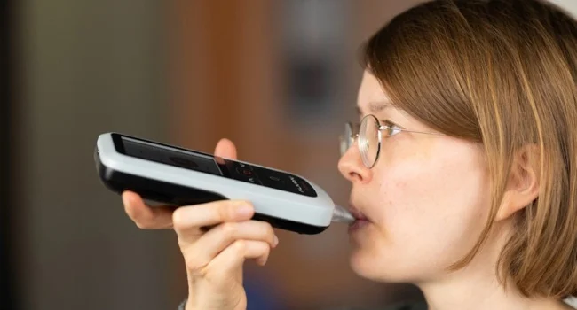
Noninvasive analysis of exhaled air can help doctors determine whether lung nodules are malignant or benign. According to research conducted by Australian scientists, this approach can contribute to the early diagnosis of lung cancer and reduce the need for invasive interventions. The researchers analyzed exhaled air using two complementary methods. The first is the electronic nose (eNose), which detects the characteristic chemical patterns of exhaled air. The second is gas chromatography with mass spectrometry (GC-MS), which detects certain volatile organic compounds. Both methods were significantly better at distinguishing between malignant and benign pulmonary nodules than models based on standard clinical data alone, said Griffith University's Institute of Biomedicine and Glycomics professor and MedComm. Chamindi Punyadira, author of the study published in the journal. Currently, invasive methods are used to evaluate lung nodules, including CT-guided biopsy and bronchoscopy. In some cases, surgery may be necessary to determine the nature of the suspicious growth. According to researchers, about 2.5 million new cases of lung cancer are diagnosed worldwide every year. Study co-author Zhang Xi from Griffith University noted that performing lung nodule biopsies can be technically challenging, especially if the nodules are small or in a hard-to-reach location. Intervention can also � lead to complications, including pneumothorax, an accumulation of air in the pleural cavity that can cause lung arrest. At the same time, the researchers stressed that breath analysis is not intended to replace a biopsy. It is It can help doctors identify patients whose nodules are more likely to be malignant and prioritize invasive testing for those who need it. According to scientists, it is a non-invasive test Adoption into standard clinical practice may also further reduce the number of unnecessary invasive procedures for patients with benign nodules.
Society
Exhaled air analysis can help distinguish cancerous lung nodules from benign ones. research
Source: KARTSIQ